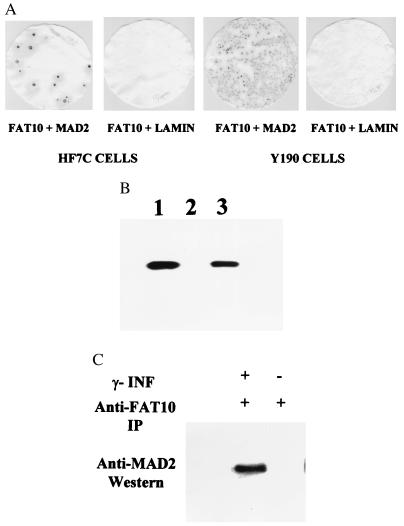
Figure 5.
The specific interaction of FAT10 with MAD2 in a yeast two-hybrid screen (A), in vitro on a GST column (B), and in vivo in immunoprecipitates (C). (A) The β-galactosidase assays show positive (blue) for specific interaction between FAT10 and MAD2 but negative (white) for FAT10 and human lamin C. (B) The FAT10-GST fusion proteins were produced with the GST expression vector, pGEX-4T-1 (Pharmacia). The 35S-labeled MAD2 was added to the GST column in RPMI 1640 medium. The 35S-labeled MAD2 retained on GST columns was washed, eluted, and analyzed in 10% SDS/PAGE. (1) 35S- labeled MAD2 control, (2) 35S MAD2 eluted from GST protein bound GST column, (3) 35S MAD2 from FAT10-GST bound GST column. (C) Anti-FAT10 polyclonal antibody was used to immunoprecipitate FAT10 in the JY cell lysates in 50 mM Tris (pH 8)/150 mM NaCl/1% Triton X-100/100 μg/ml PMSF and a protease inhibitor mixture (Boehringer Mannheim). The antibody–FAT10 complex was pulled down with protein A Sepharose 6B (Pharmacia) resin. After extensive wash, the final immunoprecipitate was analyzed by Western blot with anti-MAD2 antibody (Santa Cruz Biotechnology). The MAD2 clearly coprecipitates with FAT10 in the γ-IFN induced JY cell lysate but not in the uninduced JY cell lysate.

