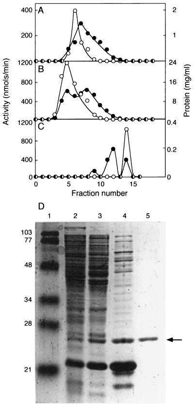
Figure 1.
Purification of RhlI from clarified cell extracts. (A) HiTrap Q column chromatography. (B) HiTrap S column chromatography. (C) Superdex 75 column chromatography. ○, RhlI activity (nmol of butyryl-HSL produced min−1 in each fraction); ●, protein levels. (D) SDS/PAGE of RhlI activity peaks. Lane 1, molecular mass standards (prestained low-range markers, Bio-Rad); lane 2, 30 μg of cell extract; lane 3, 35 μg of protein from pooled HiTrap Q fractions 3–8; lane 4, 30 μg of protein from pooled HiTrap S fractions 5–8; lane 5, 1.4 μg of protein from pooled Superdex 75 fractions 13–15. The numbers to the left indicate the molecular mass of the standard. The arrow indicates RhlI.

