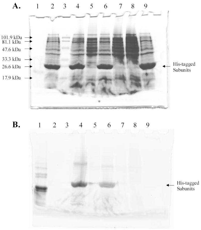
Figure 1.

Analysis of His-tagged R-PE apo-subunits and holo-subunits by SDS-PAGE. A, Detection of proteins by Coomassie staining. B, Detection of proteins by fluorescence imaging (excitation wavelength 532 nm; emission filter 580BP30). Lane 1, R-PE subunits; lane 2, lysate of cells that expressed apo-alpha subunit of R-PE; lane 3, prestained MW standards, low range 18–106 kDa (Bio-Rad); lane 4, lysate of cells that expressed apo-alpha subunit of R-PE and were incubated with PEB. Holo-alpha subunit was formed; lane 5, lysate of cells that were not induced for expression of apo-alpha subunit of R-PE but were incubated with PEB; lane 6, lysate of cells that expressed apo-beta subunit of R-PE and were incubated with PEB. Holo-beta subunit was formed; lane 7, lysate of cells that were not induced for expression of apo-beta subunit of R-PE but were incubated with PEB; lane 8, lysate of BL21(DE3) cells. Cells were incubated with PEB, but neither contained plasmids bearing subunit genes nor were treated with IPTG; lane 9, lysate of cells that expressed apo-beta subunit of R-PE.
