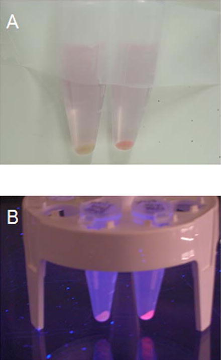
Supplementary Figure 3.

MBP-R-PE subunit solutions after attachment of PEB compared to solution of free PEB. Vials from left to right contained the following solutions: 1. cytoplasmic MBP-R-PE alpha subunit and PEB in maltose elution buffer, 2. PEB in maltose elution buffer. 3. cytoplasmic MBP-R-PE beta subunit fusion and PEB in maltose elution buffer, A, Daylight color of solutions. Solutions are pink in color due to the same color of free PEB and PEB bound to fusion proteins. B, Color of solutions under UV illumination. Orange fluorescence from solutions containing fluorescent subunit fusions is seen. These images demonstrate the large increase in fluorescence of PEB chromophore after attachment to soluble apo-subunit fusion proteins.
