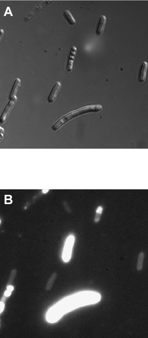
Figure 6.

Subcellular localization of fusion protein consisting of periplasmic MBP and R-PE apo-alpha subunit after incubation of cells with PEB. Elongated cells were much brighter than normal cells probably because they expressed more of the fusion protein. Fluorescent fusions were localized throughout cells in normal cells indicating that fusion proteins are soluble in the cell cytoplasm, and probably partially exported to periplasmic space. In elongated cells, fluorescent fusions were localized both in cell cytoplasm and at cell poles revealing the presence of both soluble and insoluble fusion proteins. Elongation of cells was not noticed in control cells and fluorescence of these cells was less bright than fluorescence of induced cells (images not shown).
