
Figure 2. Expression of MsMyoXVIIIAn.
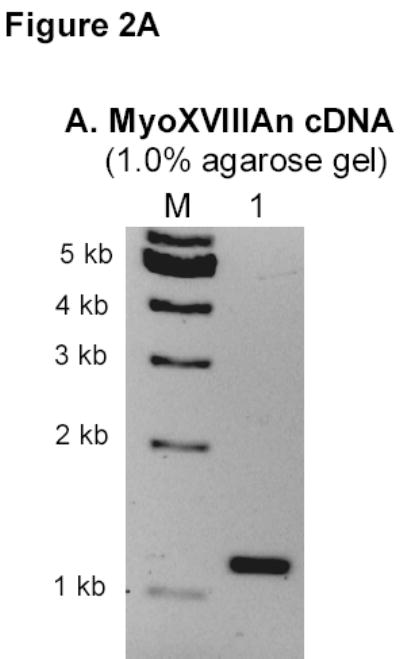
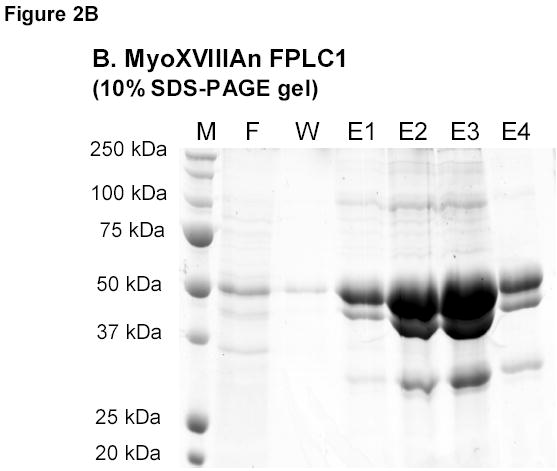
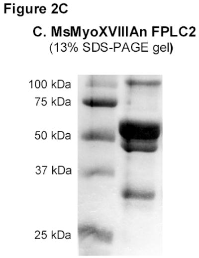
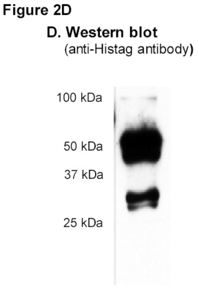
A) RT-PCR amplification of MsMyoXVIIIAn. B) Affinity chromatography of MsMyoXVIIIAn purification. Protein was separated on 10% SDS-PAGE using 10 μl FT (F), wash (W), and elution fractions (E1–E4) and stained with colloidal blue. C) Composition of 10 μg MsMyoXVIIIAn after rechromatography of E1–E4 (Figure 2B) on HisTrap column visualized on 13% SDS-PAGE with colloidal blue. D) Western blotting of 0.1 μg MsMyoXVIIIAn with anti-HisTag antibody.