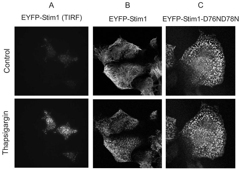
Figure 6.
Cellular movements of the Ca2+ sensor, Stim1. A: A field of several HEK293 cells expressing EYFP-Stim1 was imaged by TIRFM just prior to (upper panel) and 6.5 min after (lower panel) depletion of intracellular Ca2+ stores with thapsigargin (2 μM) in nominally Ca2+ free extracellular medium (n = greater than 10 coverslips). B,C: A field of several HEK293 cells expressing EYFP-Stim1 (B) or EYFP-Stim1-D76N, D78N (C) was imaged by fluorescence confocal microscopy just prior to (upper panel) and 10 min after (lower panel) depletion of intracellular Ca2+ stores with thapsigargin (2 μM) in HBSS containing 1.8 mM Ca2+. For B and C, data are representative of 3 independent experiments.

