
Fig. 1.
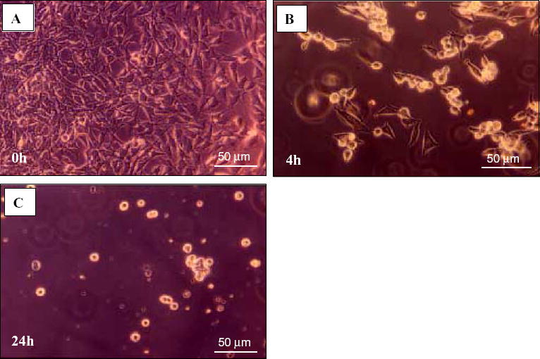
Representative photos of the C17.2 neural stem cell line during incubation with CSF from a diseased MRL-lpr mouse. A) The cell layer was confluent at 0 h, i.e. before CSF was added. B) Adhered cells lost their processes and became rounded within 4 h, detached from surface, and C) most of them degenerated within 24 h.