
Fig. 6.
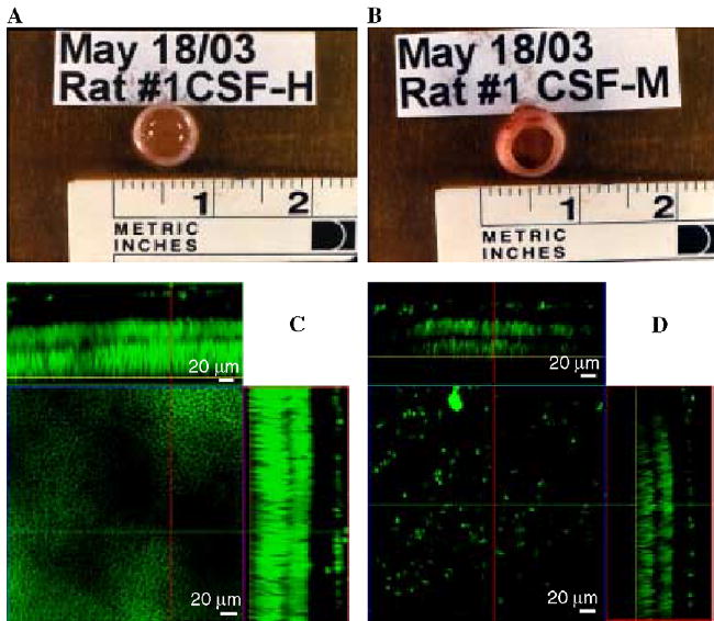
Eyes extracted from a rat four days after a coded intravitreal injection (one eye each) of 5 μl of CSF collected from control MRL+/+ (A) or diseased MRL-lpr mouse (B). Necrotic changes were detected in 7 out of 10 rats after administration of CSF from diseased MRL-lpr mice. Images of retinas exposed to CSF from MRL+/+ (C) and MRL-lpr mouse (D), viewed under confocal microscopy. Necrotic changes were detected in 7 out of 10 rats after administration of CSF from diseased MRL-lpr mice. The panel above the main square represents the ‘‘X’’ plane of the optically reconstructed section of the retina, whereas the panel on the right represents the ‘‘Y’’ plane of the section. The center squares display transverse planes within the distal photoreceptor nuclear layer (yellow line in the optical sections) of the two treatment groups. The retina treated with CSF from a MRL-lpr mouse showed severe thinning of the overall retinal thickness, as well as that of the individual layers.