
Figure 4.
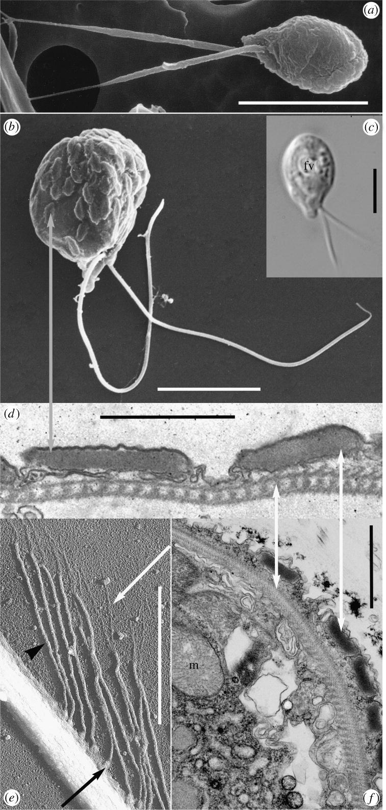
Morphology and ultrastructure of Telonema. (a–c) Whole cell: (a) Telonema subtilis scanning electron micrograph from natural sample (Gulf of Naples); (b) cultured T. antarcticum from the Oslofjord, showing cortical alveoli (grey arrow); (c) light micrograph of cultured cell (RCC 404 from Roscoff), fv, food vacuole. (d–f) Sub-cellular components of T. antarcticum: (d) section through peripheral vacuoles and cytoskeleton (white arrows); (e) detail of flagellum with flagellar tubular tripartite hairs as revealed by shadow cast whole-mount (see white arrow: distal filament; arrowhead: shaft; black arrow: base); (f) longitudinal section of embedded T. antarcticum, showing the cortical alveoli-like peripheral vacuoles, complex cytoskeleton (white arrows), m, mitochondrion with tubular crista. (a–c) Scale bar, 5 μm; (d–f) scale bar, 1 μm.