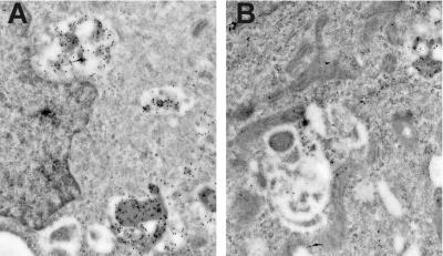
Figure 4.
Effect of tamoxifen on DAMP labeling of MCF-7/ADR cells. Acidification of the lysosomes was probed with the weak base DAMP, which accumulates in acidic organelles. (A) Electron micrograph of mouse anti-DNP and gold-conjugated anti-mouse antibodies in MCF-7/ADR cells. The gold particles indicate accumulation of DAMP within cytoplasmic organelles. The average density of gold particles was 7.02/μm2 of lysosomal area. (B) Cells incubated with tamoxifen had a substantial reduction of anti-DAMP labeling to 2.0 gold particles/μm2 of lysosomal area. Cells were incubated with DAMP, then fixed and prepared for immunoelectron microscopy as described in Materials and Methods. (Bar is 1 μm.)

