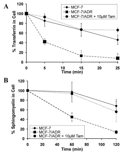
Figure 5.
Effect of tamoxifen on transport in MCF-7/ADR cells. (A) Kinetics of transport of BODIPY-transferrin to the surface. The cell-associated BODIPY-transferrin was quantified at various time points by using confocal microscopy. After 5 min only 50% of the transferrin was still associated with the MCF-7/ADR cells (■). In contrast, more than 90% remained with MCF-7/ADR cells that had been incubated with 10 μM tamoxifen (●). After 25 min less than 10% of the transferrin remained with the control MCF-7/ADR cells and more than 60% remained with the tamoxifen-treated cells. The rate of transferrin transport in MCF-7/ADR cells treated with tamoxifen was similar to the rate in drug-sensitive MCF-7 cells (⧫). (B) Kinetics of transport of BODIPY-sphingomyelin to the surface. The kinetics of transport of the lipid sphingomyelin from the TGN to the surface was quantified as described in Materials and Methods. Two hours after removal of the BODIPY-ceramide, the fluorescence in the MCF-7/ADR cells decreased to almost 20% (■). In the presence of tamoxifen (10 μM) the BODIPY-fluorescence decrease was slower (●). In the MCF-7 cells (⧫) the rate of transport of the BODIPY-sphingomyelin to the surface was similar to that of the MCF-7/ADR cells with tamoxifen.

