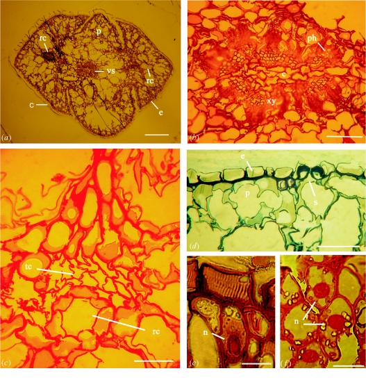
Figure 2.
Light microscope images of thin sections of the amber cypress (a–e) and of a recent Juniperus leaf (f). (a) Unstained cross-section of the entire stem and leaves, showing the cuticle (c) the epidermis (e), the parenchyma (p), the vascular system (vs) and the resin canals (rc). (b) Safranine stained vascular system with phloem (ph), xylem (xy) and core tissue (c). (c) Safranine stained tissue of a small resin canal (rc) formed by a layer of glandular cells adjacent to a layer of tracheid-like cells (tc) with double bordered pits. (d) Methylene blue stained tissue of the epidermis (e) with a stomatal pore (s) and the parenchyma (p). (e) A parenchyma cell stained with Stains All displaying a nucleus (n) and intensively stained cell walls. (f) Parenchyma cells from a leaf of a recent Juniperus sabina with nuclei (n) stained with Stains All. Size bars: (a) 300μm; (b–d) 50μm; (e, f) 20μm.

