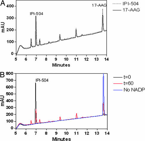
Fig. 1.
Representative HPLC chromatograms of a 60-min time point of IPI-504 or 17-AAG incubated with human liver microsomes. (A) IPI-504 (50 μg/ml) (dashed line) or 17-AAG (solid line) was incubated in human liver microsomes. The reactions were quenched at 60 min with an equal volume of 2:1 methanol:270 mM citrate (pH 3), 0.5% (wt/vol) EDTA, and 0.5% (wt/vol) ascorbate to preserve IPI-504 and analyzed by HPLC (λ = 230 nm). (B) IPI-504 (50 μg/ml) was incubated with 0.8 mg/ml human liver microsomes for 0 (black line) and 60 min (red line), as well as for 60 min without NADP as a control reaction (blue line). The reaction was quenched by addition of an equal volume of 2:1 methanol:270 mM citrate (pH 3), 0.5% (wt/vol) EDTA, and 0.5% (wt/vol) ascorbate to preserve IPI-504 and analyzed by HPLC (λ = 230 nm).

