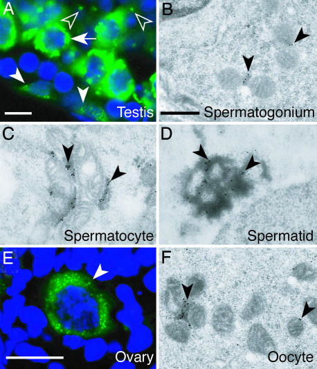
Fig. 1.
TDRD1 is a component of nuage in both male and female germ cells. (A) Immunostaining of a section of an adult testis with anti-TDRD1 antibody (green) counterstained with Hoechst dye (blue). Spermatogonia, pachytene-spermatocytes, and round spermatids are indicated by arrowheads, an arrow, and open arrowheads. (B–D) Immunoelectron microscopy of testis sections with anti-TDRD1 antibody. Intermitochondrial cement in a spermatogonium (B) and a spermatocyte (C) and a chromatoid body in a round spermatid (D) are indicated by arrowheads. (E) An adult ovary section immunostained for TDRD1 (green) and counterstained with Hoechst dye (blue). The arrowhead indicates a primary ooctye. (F) Immunoelectron microscopy of a primary oocyte for TDRD1. The arrowheads mark TDRD1 signals on intermitochondrial cement. (Scale bars: A and E, 10 μm; B–D and F, 1 μm.)

