Fig. 2.
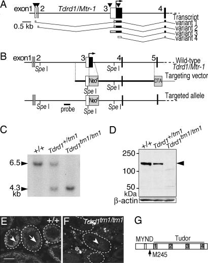
Targeted mutagenesis of Tdrd1. (A) The 5′ regions of four transcript variants of Tdrd1 are depicted under the exon structure. The arrowheads indicate transcription initiation sites, the open and filled boxes represent untranslated and translated regions, and the arrow marks the first coding ATG of transcript variants 1–3. (B) The targeted allele of Tdrd1. The neomycin-resistant cassette replaced the first coding ATG of transcript variants 1–3 and the promoter region of transcript variant 4. (C) Southern blot analysis of tail genomic DNAs digested with SpeI and hybridized with the probe shown in B. (D) Western blotting of testis lysates probed with anti-TDRD1 (Upper) and anti-β-actin (Lower) antibodies. The arrowhead indicates wild-type TDRD1. The two faint bands seen in the Tdrd1tm1/tm1 lane are nonspecific signals, because they are not detected in the Tdrd1tm1/+ lane. (E and F) Immunostaining of sections of wild-type (E) and Tdrd1tm1/tm1 (F) testes with anti-TDRD1 antibody. Spermatocytes and round spermatids are indicated by arrowheads and arrows. The dotted lines demarcate seminiferous tubules, and autofluorescence is labeled with an asterisk. (G) The domain composition of wild-type TDRD1. A potential first ATG in the truncated Tdrd1tm1 mRNAs (see text) corresponds to methionine 245 of the wild-type TDRD1 (arrow). (Scale bar: 100 μm.)

