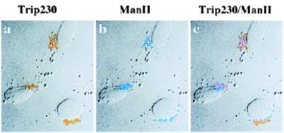
Figure 2.
Colocalization of Trip230 and mannosidase II to the region of the Golgi complex. Cells were fixed and double-immunostained with anti-Trip230 and antimannosidase II antisera. Shown in this representative field is the indirect immunofluorescence antibody staining imaged by laser-scanning confocal microscopy. a, pattern of immunofluorescence using anti-Trip230 polyclonal serum (1:1,000 dilution) and FITC-tagged anti-mouse IgG secondary antibodies that localize Trip230 to perinuclear cytoplasm (pseudocolored orange); b, rabbit antimannosidase II (a Golgi enzyme) antiserum and Texas red-tagged anti-rabbit secondary antibody labeling of the Golgi complex (pseudocolored blue); c, digital overlay of anti-Trip230 and mannosidase II antiserum images showing that while in the same vicinity of the Golgi, the overlap of Trip230 immunofluorescence is incomplete.

