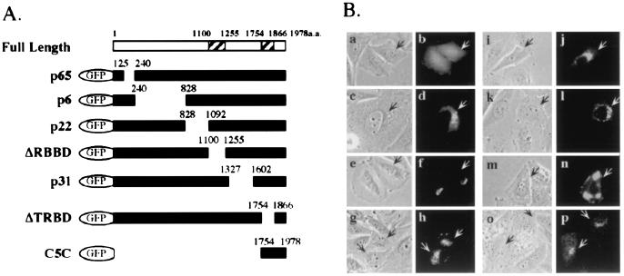
Figure 3.
Mapping of Trip230 Golgi-localization domain. (A) Diagram of serial deletion constructs of GFP-Trip230 fusion proteins with the residues that were deleted indicated above the solid bars. ΔRBBD indicates deletion of the RB-binding domain (amino acids 1100–1255); ΔTRBD indicates deletion at TR-binding domain (amino acids 1754–1866). C5C shows the GFP fusion containing Trip230 residues 1754–1978 fused to GFP. (B) Localization of GFP and GFP-Trip230 fusion proteins in transiently transfected Saos-2 cells. Phase-contrast images (a, c, e, g, i, k, m, and o) show cell morphology for each transfection. GFP autofluorescence (green, b, d, f, h, j, l, n, and p) shows the subcellular location of the various GFP-Trip230 fusion proteins. a and b, fluorescence of GFP alone; c and d, p65; e and f; p6; g and h; p22; i and j; ΔRBBD; k and l; p31; m and n; ΔTRBD; and o and p, C5C. Arrows indicate transfected cells.

