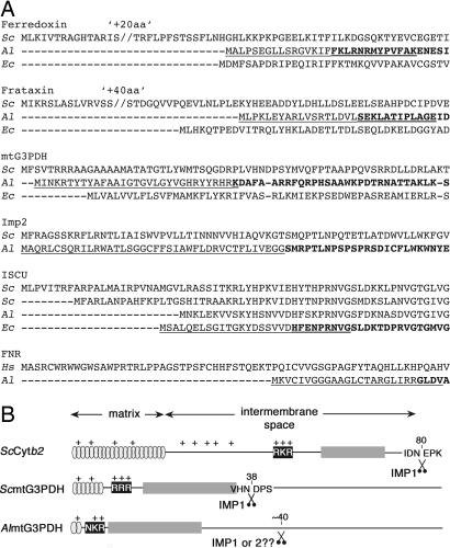
Fig. 2.
N-terminal regions of microsporidian mitosomal proteins. (A) A. locustae and E. cuniculi mitosomal proteins in comparison with their yeast (S. cerevisiae) or Homo sapiens homologues. N-terminally truncated versions used in Fig. 3B are indicated by bold. N-terminal signals fused to GFP are underlined. (B) Schematic representation of the presequence of AlmtG3PDH in comparison to yeast ScCytb2 and ScmtG3PDH with regions corresponding to a basic amphipathic helix containing the matrix targeting information (gray) and the hydrophobic intermembrane sorting sequence (dark gray) preceded by a cluster of positively charged residues (black) important for recognition of the sorting sequence. For AlG3PDH a possible processing site is indicated around residue 40, an estimate from deletion experiments (see Fig. 4C).

