
Figure 1.
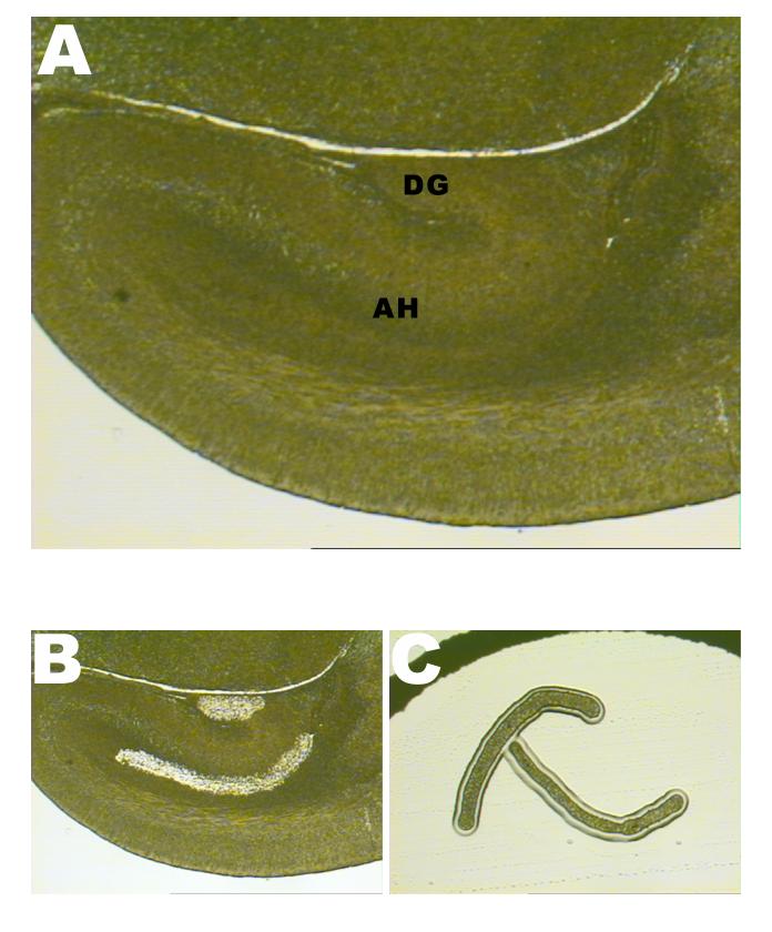
Selected hippocampal areas for laser capture microdissection.
- A general view of the E17 mouse fetal hippocampus. AH denotes the Ammon’s horn ventricular and subventricular zones; DG denotes the prime germinal zone of the dentate gyrus.
- The same section of the hippocampus after cell removal using the Laser Capture Microdissection (LCM) technique.
- Two captured areas of Ammon’s horn ventricular and subventricular zones (right and left, from the same paraffin-embedded section), used for subsequent DNA extraction and bisulfite sequencing.