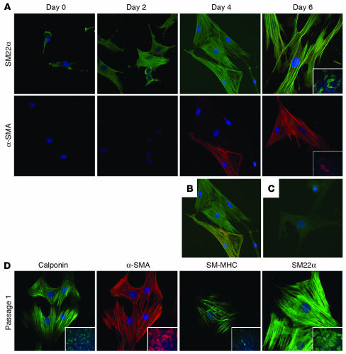
Figure 3. SMC-specific proteins are expressed in SD-MAPCs differentiated with TGF-β1.
(A) Temporal characterization of SM22α and α-SMA expression on days 0, 2, 4, and 6 (magnification, ×40). SM22α was detected at low levels on day 0, which increased and localized to stress fibers as early as day 2. α-SMA expression was absent on day 0 and detected by day 4. (B) Colocalization of SM22α (green) and α-SMA (red) further demonstrates that expression of SM22α precedes that of α-SMA. (C) IgG control. (D) Expression of the SMC-specific proteins calponin, α-SMA, SM-MHC, and SM22α in SD-MAPC-SMCs passaged once in expansion medium. Representative example of more than 5 experiments; insets: magnificiation, ×10.

