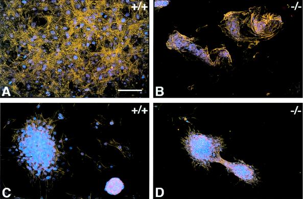
Figure 2.
Mouse skin fibroblasts cultured on silicone rubber disks for 48 h. Control (A and C) and TSP2-null fibroblasts (B and D) were plated on PDMS (A and B) and ox-PDMS (C and D). Cells were visualized by staining of their actin cytoskeleton with phalloidin (orange) and their nuclei with 4′,6′-diamidino-2-phenylindole (DAPI) (blue). Control fibroblasts formed a confluent overlapping monolayer on PDMS (A). On ox-PDMS, the monolayer was loosely attached and was disrupted and reassembled into aggregates during the manipulations associated with culture (C). TSP2-null fibroblasts failed to form a monolayer on PDMS (B) or on ox-PDMS (D) but were associated in aggregates. On ox-PDMS the cellular aggregates tended to slide on the biomaterial surface with movement of the culture medium. (Bar = 50 μm for A–D.)

