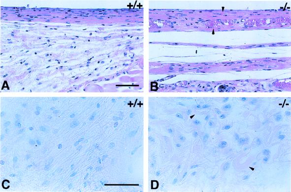
Figure 3.
FBR to implanted PDMS disks. Six-millimeter disks were implanted in control (A and C) and TSP2-null mice (B and D) and were removed 4 weeks later. Sections were stained with hematoxylin and eosin (A and B) or Verhoeff-van Gieson stain (C and D). Both treatments stain nuclei dark (blue) and cytoplasm and collagen fibers (pink). Capsules forming around the silicone disks were thicker and more highly vascularized (arrowheads) in TSP2-null mice (B). Collagen fibers in the capsules of control mice appeared dense, parallel, and organized (C). Collagen fibers in the capsules from TSP2-null mice appeared irregular in shape (arrowheads) and were less organized with respect to the implant surface (D). (Bars: A and B = 50 μm; C and D = 50 μm.)

