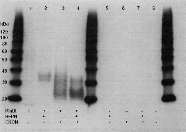
FIG. 1.

Western blot of PlnDI. Expression of PlnDI in EBNA cells, purification, Western blotting, and enzyme digestions were performed as described in Materials and Methods. Lane 1, undigested PlnDI; lane 2, PlnDI digested with heparinases I, II, and III (HEPN); lane 3, PlnDI digested with chondroitinase AC (CHON); lane 4, PlnDI digested with both HEPN and CHON; lanes 5-8, negative controls in which PlnDI was omitted. Numbers in the margin denote the migration position of protein molecular weight markers (in kDa).
