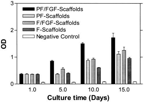
FIG. 8.

hBMSC proliferation in PlnDI-coated collagen I scaffolds. hBMSCs derived from human bone marrow were expanded as described in Materials and Methods. Collagen I fibril scaffolds were seeded with an hBMSC suspension (2 ×10⁵/mL) in DMEM supplemented with 10% (v/v) FCS. Cell-seeded scaffolds then were cultured as described in Materials and Methods. Cell-free collagen I scaffolds served as a negative control. Scaffolds were collected on the indicated days postseeding and cell proliferation was determined by BrdU incorporation assay as described in Materials and Methods. The results reflect means ± SD of triplicate determinations in each case.
