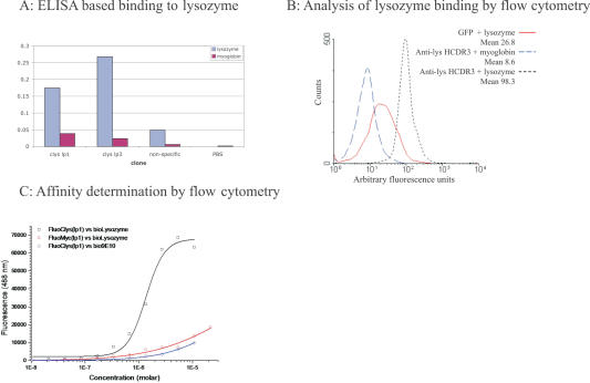
Figure 2.
(A) ELISA was carried out by first interacting biotinylated antigen with the GFP clone of interest and then capturing on a neutravidin coated plate. GFP binding was revealed using SV5, a monoclonal antibody (73), which recognizes a tag appended to the C-terminus. The non-specific clone was GFP containing the myc epitope (74) at the loop 3 position, and indicates the level of binding due to a similarly disrupted GFP molecule. clyslp1 and clyslp3 have the lysozyme binding HCDR3 inserted into loop 1 and loop 3, respectively. (B) Streptavidin coated beads were incubated with either biotinylated lysozyme or myoglobin, (which serves as the negative control for non-specific binding to coupled beads) and subsequently with GFP or GFP containing the anti-lysozyme HCDR3 inserted at loop 1 (c-lys1). Analysis was carried out using a FACSCalibur. (C) As in (B), except that different concentrations of c-lys1 were used and the mean fluorescence of the non-specific (myoglobin) beads was subtracted from the fluorescence of the lysozyme coated beads and plotted. The affinity is calculated from the concentration of c-lys1, which gives half maximal fluorescence.

