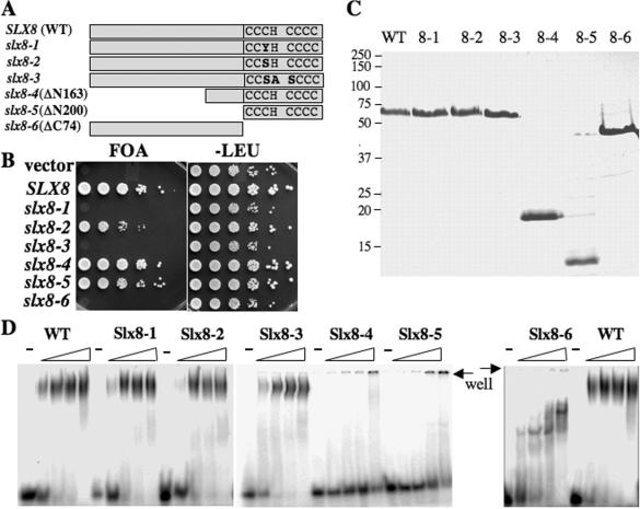
Figure 5.
Structure–function analysis of Slx5–Slx8. (A) Schematic diagram of the wild-type and mutant Slx8 proteins indicating the conserved RING-finger residues. (B) Strain VCY1525 (sgs1Δ slx8Δ), which contains the complementing plasmid pJM500 (SGS1/URA3), was first transformed with centromere-based vector (pRS415) or vector containing the indicated SLX8 allele. Complementation of slx8Δ phenotype was then tested by serial dilution and spotting onto media containing 5-FOA to select against pJM500 or onto media lacking leucine as control. (C) An aliquot of 2 μg of the indicated Slx8 protein was resolved by SDS–PAGE and stained with Coomassie blue. (D) The indicated Slx8 protein (0, 0.4, 1.2 and 4 pmol) was incubated with 5 fmol of the 45 bp 5′-32P-labeled substrate and analyzed using EMSA as in Figure 2.

