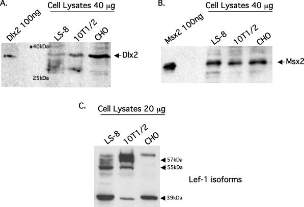
Figure 2.
Endogenous expression of Dlx2, Msx2 and Lef-1 in three cell lines. (A) Western blot of endogenous Dlx2 protein in the LS-8 tooth epithelial cell line, C3H10T1/2 pluripotent cell line, CHO cell line using the Dlx2 antibody. Whole cell lysates from each cell line were prepared and 40 μg of protein run on a 10% SDS–polyacrylamide gel. The proteins were visualized using ECL reagents from Amersham. Pure Dlx2 protein was used as a control at 100 ng and two molecular weight markers are noted (40 and 25 kDa). (B) Western blot of endogenous Msx2 protein from the same cell lines and experimental procedure as in (A). Pure Msx2 protein was used as a control at 100 ng. (C) Western blot of endogenous Lef-1 protein from the same cell lines and experimental procedures as in (A). The approximate molecular weights of the Lef-1 isoforms are noted.

