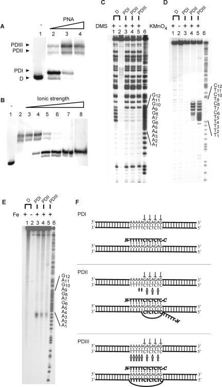
Figure 2.
Binding of PNA dodecamer to dsDNA (PD12). Autoradiographs showing binding of the indicated PNAs to dsDNA as investigated by gel-shift analysis (A and B), in situ chemical probing (C and D) and affinity cleavage (E). (A) Titration using the following PNA1708 concentrations: lane 1, w/o; lane 2, 0.25 μM; lane 3, 2 μM; and lane 4, 6 μM. Free dsDNA (D) and the formed complexes PDI12, PDII12 and PDIII12 are indicated. Analogous PD12 complexes were obtained using NTA-conjugated PNA2046 (Supplementary Figure S1). (B) PD12 complex formation as a function of ionic strength using 6 μM PNA1708 and supplemented with the following amounts of KCl: lane 1, w/o; lane 2, 5 mM; lane 3, 10 mM; lane 4, 20 mM; lane 5, 40 mM; lane 6, 80 mM; lane 7, 160 mM; and lane 8, 320 mM. (C) DMS probing of gel-purified PD12 complexes involving PNA2046: lane 1, dsDNA + DMS; lane 2, dsDNA, w/o DMS; lane 3, PDI12 + DMS; lane 4, PDII12 + DMS; lane 5, PDIII12 + DMS; and lane 6, A/G sequence. (D) Permanganate probing of gel-purified PD12 complexes involving PNA2046: lane 1, dsDNA + KMnO4; lane 2, dsDNA, w/o KMnO4; lane 3, PDI12 + KMnO4; lane 4, PDII12 + KMnO4; lane 5, PDIII12 + KMnO4; and lane 6, A/G sequence. (E) Fe/NTA affinity cleavage of gel-purified PD12 complexes involving PNA2046: lane 1, dsDNA + Fe/NTA; lane 2, dsDNA, w/o Fe/NTA; lane 3, PDI12 + Fe/NTA; lane 4, PDII12 + Fe/NTA; lane 5, PDIII12 + Fe/NTA; and lane 6, A/G sequence marker. (F) Structural assignment as based on the probing results [arrow (KMnO4 hypersensitivity), perpendicular symbol (DMS protection)]. The 168 bp XbaI–PvuII restriction fragment of p322 was used either 5′-kinase labelled (A–C, E) or 3′-Klenow labelled (D) with 32P-phosphate at the XbaI site. PNA incubations were at pH 6.5 as described in Materials and Methods.

