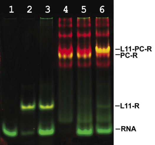
Figure 12.
Stimulation of the L10/L124–RNA interaction in the presence of L11 analyzed by non-denaturing PAGE with two color fluorescent staining. The protein component is visualized with SYPRO® Red Protein Gel Stain (red) and the RNA component with SYBR® Green I (green). The yellow color represents the protein–RNA complexes because of the superimposed green and red signals from the RNA (SYBR Green I) and protein (SYPRO Red) channel. Lane 1, RNA fragment Mja23S-95; lanes 2, 3, MjaL11 mixed with RNA fragment in a molar ratio of 1:1, 1:2 respectively; lanes 4 and 5, MvaL10/L124 mixed with RNA fragment in a molar ratio of 4:1, 2:1, respectively; lane 6, MvaL10/L124 mixed with RNA fragment and L11 r-protein in a molar ratio of 2:1:1. RNA, RNA fragment Mja23S-95; PC-R, L10/L124 bound to RNA; L11-R, L11 bound to RNA; L11-PC-R, complex of L11 and L10/L124 with RNA.

