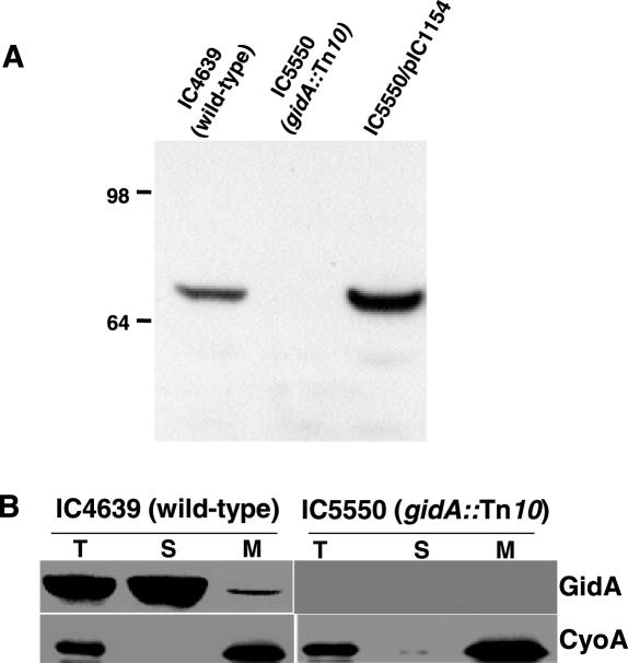
Figure 2.
(A) Specificity of the anti-GidA antibody. Equal amounts of total extracts of strains IC4639, IC5550 and IC5550 harbouring plasmid pIC1154 (where gidA is under control of Ptac promoter and LacI repressor) were subjected to western blot analysis using anti-GidA antibody. Strain IC5550/pIC1154 was grown in the absence of inducer IPTG, but there is escape of gidA expression. Molecular weight markers (on the left) are in kDa. (B) Subcellular localization of GidA. Western blot analysis with anti-GidA and anti-CyoA antibodies of cell fractions from strains IC4639 and IC5550 is shown. Equal amounts of bulk protein (50 μg) were loaded in each lane. T, total cell lysate; S, soluble fraction; M: membrane fraction.

