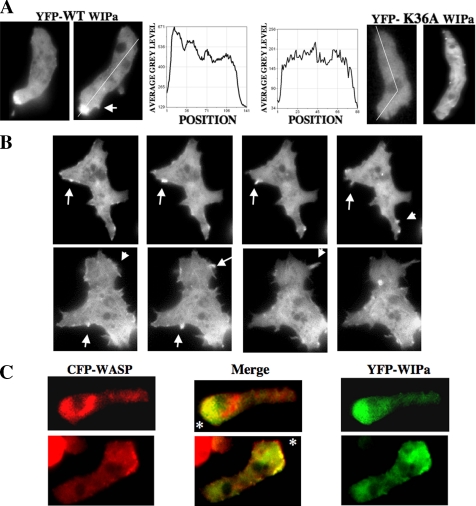
Figure 3.
WIPa localizes to leading edge, sites of membrane protrusion, and colocalizes with WASP. (A) WIPa localization in chemotaxing cells. YFP-tagged WIPa, but not actin-binding domain mutant K36A. WIPa, is enriched at the leading edge of polarized cells. (B) Transient WIPa localization at sites of membrane protrusion. Images were taken at 6-s intervals. (C) YFP-tagged WIPa colocalizes with CFP-tagged WASP at the leading edge of chemotaxing cells. Asterisk indicates location of the micropipette tip releasing 100 μM cAMP.

