Figure 5.
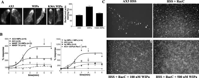
WIPa stimulates de novo F-actin polymerization. (A) Barbed-end staining of cells expressing wild-type WIPa or mutant (K36A WIPa). At least 60 cells taken from three independent experiments were used for quantitation. (B) In vitro F-actin polymerization assay. Supernatants of either wild-type AX3, WASPTK, or hp WIPa cells were stimulated with GTPγS-bound RacC at time 0 (or with GDPβS-bound RacC as indicated ◇). WIPa, 100 nM, was added 15–30 s after RacC addition where indicated. Reactions were stopped at the indicated time points with actin buffer containing TRITC-phalloidin, and F-actin was pelleted by centrifugation. The amount of F-actin at time 0 was standardized as 100%. (C) Visualization of polymerized F-actin. Supernatants were incubated with or without 100 nM GTPγS-charged RacC or WIPa for 5 min on a poly-l-lysine–treated glass coverslip and then fixed.

