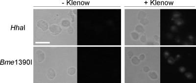
Figure 3.
Bright field and epifluorescence images of in situ ligation-stained cells. Exponentially growing cells were treated with HhaI (1-base pair 3′ overhang) and Bme1390I (1-base pair 5′ overhang) endonucleases. The cells in the right column were treated with Klenow (+ Klenow, HhaI) or Klenow + dNTPs (+ Klenow, Bme1390I) before in situ ligation staining. Left, phase-contrast microscopy; right, fluorescence microscopy of the same cells. Bar, 5 μm.

