Figure 6.
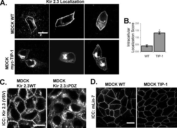
Kir 2.3 is mislocalized to a vesicular compartment in MDCK/TIP-1 cells. (A) Confluent monolayers of wild-type (WT, top panels) or MDCK cells stably transfected with myc-TIP-1 (MDCK myc-TIP-1, bottom panels), were transiently transfected with VSV-Kir 2.3 and stained with anti-VSV antibody. Images were chosen to document the full spectrum of phenotypes observed for each cell type. Scale bar, 10 μm. (B) Cells were scored for intracellular localization of Kir 2.3 (2, strong; 1, moderate; 0, none) by a blinded observer. Data are reported as the average Kir 2.3 intracellular localization score for cells from three separate infections (n = 100; p < 0.001). (C) MDCK cells stably expressing a mutant Kir 2.3 protein lacking the PDZ ligand (ΔPDZ) display a similar mislocalization phenotype as caused by TIP-1 expression. Cells are labeled with anti-VSV antibodies, detecting Kir 2.3. (D) MDCK WT and myc-TIP-1 cells were stained with anti-mLin-7 antibodies for endogenous mLin-7. Scale bar, 20 μm.

