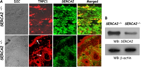
Figure 2.
SERCA2+/− mice exhibit increased TRPC1 expression. (A) Immunofluorescence in skin sections (10 μm) obtained from SERCA2+/+ or SERCA2+/− mice using anti-TRPC1 and anti-SERCA2 antibodies. Fluorescently labeled secondary antibodies were used to label TRPC1 and SERCA2 proteins. Images were taken using 63× objective. (B) Western blots performed on samples obtained from SERCA2+/+ and SERCA2+/− mice and probed using SERCA2 and β-actin antibodies.

