Figure 7.
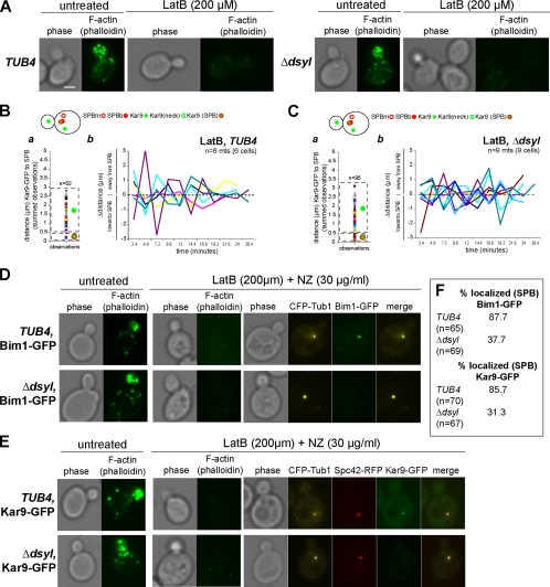
Stable localization of Kar9 in the bud depends on cortical actin. (A) Actin staining of TUB4 and tub4-Δdsyl cells with and without treatment of 200 μM LatB. Treatment with LatB resulted in the depolymerization of the actin cytoskeleton in TUB4 and tub4-Δdsyl cells. Respective phase images are shown. Bar, 2 μm. (B, a) In wild-type cells treated with LatB, Kar9–GFP foci distributed to at the bud cortex as well as the SPB. After latrunculin treatment, microtubules (Kar9–GFP foci) in wild-type cells were highly dynamic. In tub4-Δdsyl cells treated with latrunculin, the distribution of Kar9–GFP foci relative to the SPB were rescued to near wild-type levels (C, a). Kar9 dynamics were also rescued to near wild-type levels (C, b). (D–F) Analysis of Bim1 and Kar9 SPB localization in cells treated with 200 μM LatB (to disrupt F-actin) and 30 μg/ml NZ (to depolymerize astral microtubules). CFP–Tub1 was used to visualize the collapsed spindle and Alexa 488-phallodin to confirm the disruption of the actin cytoskeleton in the presence or absence of latrunculin and NZ. The majority of wild-type cells (87.7%; n = 65) treated with LatB and NZ had detectable Bim1–GFP associated with the collapsed spindle (D and F). In contrast, the percentage of LatB and NZ treated tub4-Δdsyl cells with detectable Bim1–GFP at SPBs was reduced (37.7%; n = 69) (D and F). Similarly, the localization of Kar9–GFP in tub4-Δdsyl cells treated with LatB and NZ was reduced; 31.3% of tub4-Δdsyl cells (n = 67) had detectable Kar9–GFP compared with 78% of wild-type cells (n = 70; E and F).

