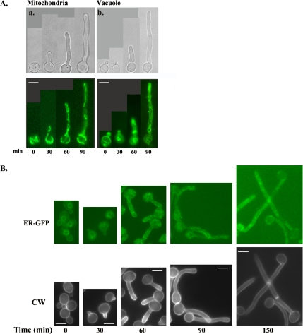
Figure 2.
ER, mitochondria, and vacuoles are randomly distributed in hyphal cells. Cells (BWP17) were induced to form hyphae by growth at 37°C and the addition of serum. Aliquots of cells were removed every 30 min and processed for fluorescence microscopy. (A) Mitochondria (a) were visualized by staining cells with DiOC6. Vacuoles (b) were visualized using MDY-64. (B) ER was visualized in a strain expressing an HDEL-tagged Kar2-GFP fusion protein (ER-GFP; see description in Materials and Methods). Cells expressing ER-GFP were induced to form hyphae, removed at the indicated times, and viewed directly for GFP localization, or stained with CW to visualize cell wall and septa as described in Materials and Methods. Bars, 5 μm.

