Figure 7.
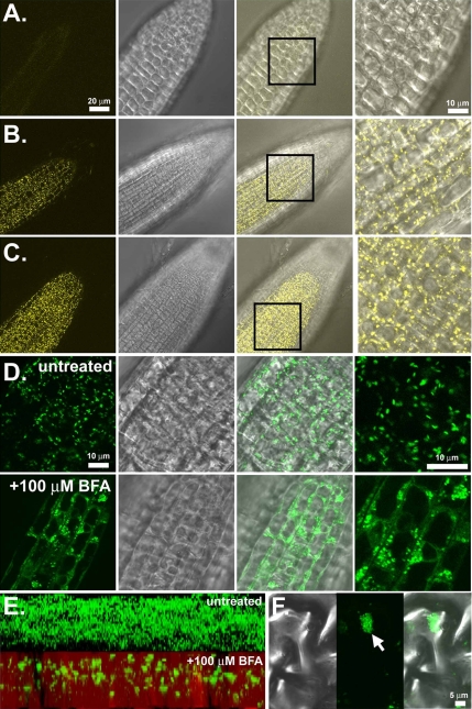
SGB1 is localized to the Golgi apparatus. (A) Wild type (Col-0) roots. (B) Imaging of SGB1 promoter-driven SGB1-YFP in the wild-type background. Individual Golgi stacks are seen as punctuate structures rapidly moving throughout the cytosol. (C) Imaging of native promoter driven SGB1-YFP in the agb1-2 background. (D) SGB1-GFP localization is sensitive to brefeldin-A (BFA). GFP imaging of the 35S promoter-driven SGB1-GFP before (untreated) and after (+100 μM BFA) treatment with BFA for 90 min, demonstrating the change in localization. (E) Z-stack reconstruction of 35S::SGB1- GFP expression in leaf tissues before (above) and after (below) 100 mM BFA treatment. (F) High-magnification imaging of a single BFA compartment in a leaf epidermal cell expressing SGB1-GFP. Left, DIC; middle, fluorescence; and right, merge.

