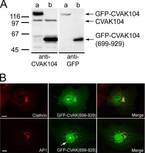
Figure 10.
Subcellular localization of GFP-CVAK104-(699-929) in COS-7 cells. (A) Western blot analysis of lysates from COS-7 cells either expressing GFP-CVAK104 full-length (a) or GFP-CVAK104-(699-929) (b). Blots are stained for CVAK104 and for GFP. (B) GFP-CVAK104-(699-929)–expressing cells were stained for clathrin HC and AP1, respectively. A blob-like structure positive for GFP-CVAK104-(699-929) that does not colocalize with AP1 is marked by an arrow. Note, that the transiently expressed GFP-CVAK104-(699-929)-fusion protein is also localized to the nucleus (denoted by asterisks). Bars, 10 μm.

