Figure 6.
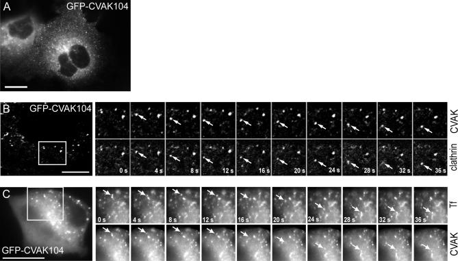
Live fluorescence microscopy of GFP-CVAK104. (A) Frame of a video (see Supplemental Movie 6A.mov) showing COS-7 cells transfected with GFP-CVAK104. (B and C) COS-7 cells were transfected with GFP-CVAK104 and either cotransfected with RFP-tagged clathrin light chains (B) or incubated with Texas Red-labeled transferrin (C) and then analyzed by live cell imaging (see Supplemental Movies 6B.mov and 6C.mov). Frames were acquired every 4 s. The series of frames are taken from the movies and show an enlarged region of the cells. The arrows indicate moving structures positive for both GFP-CVAK104 and either RFP-CLC or transferrin. The frames of the movie illustrated in B are confocal images. Bars, 10 μm.

