Figure 9.
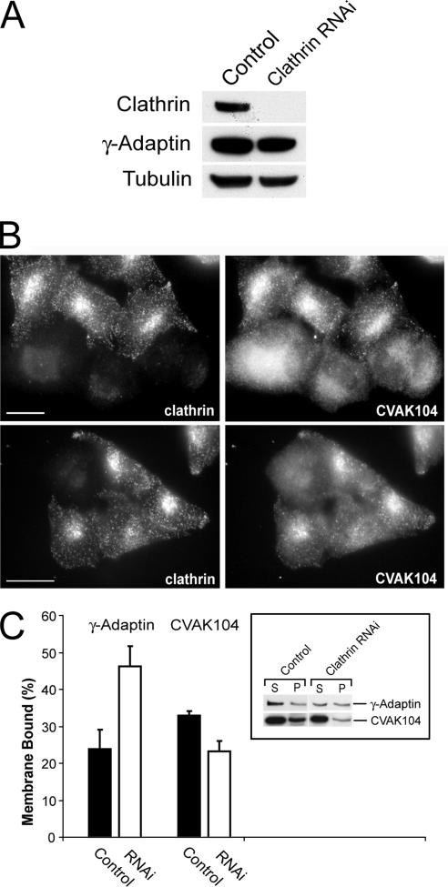
Requirement of clathrin for the subcellular localization of CVAK104. (A) Western blot analysis of lysates from clathrin siRNA- and mock-transfected HeLa cells, respectively. Blots are stained for clathrin heavy chain, the γ-adaptin subunit of AP1, and for tubulin. (B) Distribution of CVAK104 in clathrin-depleted HeLa cells. To view knockdown and control cells within the same field, control and knockdown cells were trypsinized, mixed, and replated 24 h before immunostaining with antibodies to CVAK104 and clathrin HC. In clathrin-depleted cells, CVAK104 shows a more diffuse staining. Bars, 10 μm. (C) Membrane associations of AP1 and CVAK104 upon clathrin depletion. Cells were scraped off the tissue culture plates in lyses buffer and fractionated by ultracentrifugation into membrane and cytosol fraction. Both fractions were analyzed by SDS-PAGE and Western blotting. The membrane-bound fraction of CVAK104 is reduced in clathrin knockdown cells.

