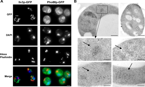
Figure 2.
Localization of actin bodies. (A) Localization of actin bodies within cells. Wild-type yeast cells expressing either Ilv3p-GFP (left) or Pho88p-GFP (right) staining the mitochondria or the endoplasmic reticulum, respectively, were grown for 7 d at 30°C in SD casa medium, fixed, and stained with DAPI and Alexa-phalloidin. In the merged bottom images, green is GFP-, blue is DAPI-, and red is Alexa-phalloidin–stained F-actin. Images are 2D maximal projection of 3D image stacks. Bar, 2 μm. (B) Immunogold localization of anti-actin antibodies linked to 10-nm gold particles on wild-type yeast cells grown for 3 d at 30°C in YPDA medium. Arrows point to clusters of gold particles. In the right top image, three clusters of gold particles within a cell are circled. Bar, 500 nm (top); 200 nm (bottom).

