Figure 5.
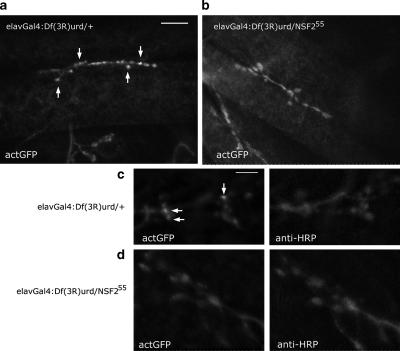
Altered actin distribution in an NSF2 loss-of-function allele. (a and b) First instar larval NMJs from Df(3R)urd/+ and Df(3R)urd/NSF255 expressing actin-GFP. Clear concentrations of the actin-GFP signal can be seen in the controls (arrows), but these are absent in the loss-of-function allele. Images were acquired at the same confocal settings. Scale bar, 10 μm. (c and d) A second pair of NMJs shown at higher magnification and double-labeled with the neural marker anti-HRP. Clear actin-GFP puncta are visible (arrows) within the Df(3R)urd/+ NMJs, whereas in the Df(3R)urd/NSF255 boutons the actin-GFP appears diffuse and the puncta are not apparent.

