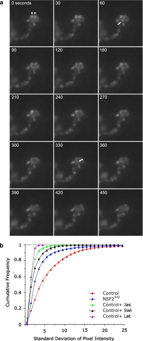
Figure 6.
Presynaptic actin is dynamic. (a) Time-lapse image sequence of actin-GFP expressed in a control nerve terminal. The time elapsed from the beginning of the sequence is indicated in the top left corner. The punctate actin structures indicated by the triangles at time zero have moved laterally by the end of the series, whereas the puncta indicated by the arrow at time 60 increases in brightness and then disappears by time 240. Finally, at time 330 some actin-GFP, at the double triangle, appears to splinter off a larger puncta and move down and out of the bouton. (b) A frequency distribution of the SD of individual pixels over the time series of image

