Figure 2.
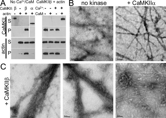
CaMKIIβ, but not α, bundles F-actin. (A) Actin was polymerized before addition of CaMKIIβ or α, as indicated. CaMKII and actin content of supernatants (S) and pellets (P) after low-speed centrifugation (10,000 × g; 20 min) were analyzed by the Western blots shown. Left, CaMKIIβ and actin were found in the pellet only when mixed; CaMKIIα and actin did not cosediment. Right, Addition of Ca2+/CaM (2 mM/3 μM), but not Ca2+ or CaM alone, prevented cosedimentation of CaMKIIβ and actin. (B) F-actin filaments are visualized by electron microscopy and do not form bundles in absence of kinase (left) or in presence of CaMKIIα (right). (C) Addition of CaMKIIβ to polymerized actin resulted in bundling of the F-actin filaments, as assessed by electron microscopy. In some areas, F-actin cross-linking without bundling was apparent (right). Bars, 50 nm.

