Figure 3.
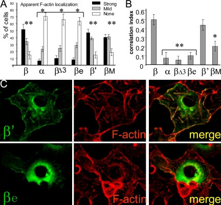
Alternative splicing modulates F-actin association of CaMKIIβ. (A) Scores of apparent F-actin localization for GFP-CaMKII variants in live Cos-7 cells. Scoring was done blind of the variant. n = 8 coverslips (50 or all transfected cells scored). There are no differences within the groups labeled * or ** (p > 0.3; analysis of variance [ANOVA]), but each * is different from each ** (p < 0.00005; two-tailed t tests) (tested were “strong” and “none” as independent values). (B) F-actin/GFP-CaMKII correlation of localization values determined using SlideBook software, after staining of F-actin with phalloidin-Texas Red as shown in C. n = 9 images. **, not different from each other (p > 0.2; ANOVA), but different from β and β′ (p < 0.00001) and βM (p < 0.05). Error bars show SEM. (C) Examples of GFP-CaMKIIβ variant localization in Cos-7 cells. F-actin was stained by phalloidin-Texas Red after fixation.

