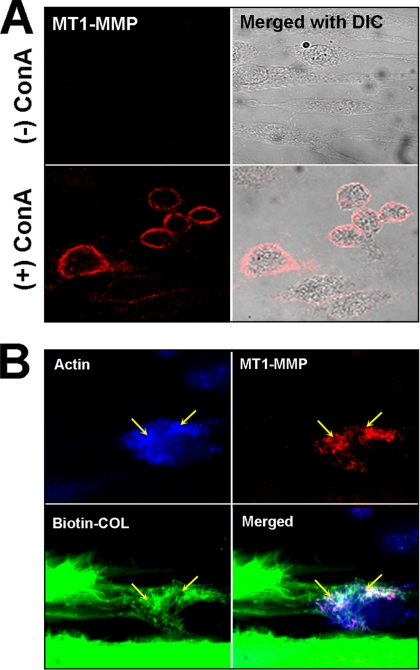
Figure 2.
Immunostaining of MT1-MMP in HGFs. (A) Laser confocal images of nonpermeabilized HGFs stained for MT1-MMP show that the enzyme (red) can be detected on the surface of cells following treatment with ConA. (B) Double immunostaining of a permeabilized, ConA-treated cell after incubation on biotinylated rat-tail tendon (Biotin-COL, green) for 72 h and stained for F-actin (blue) and MT1-MMP (red). In the merged images, the MT1-MMP can be seen to colocalize with F-actin and clusters of collagen fibrils (yellow arrows). Note that the F-actin becomes condensed after ConA treatment.

