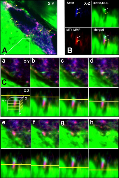
Figure 6.
MT1-MMP at sites of collagen cleavage. HGFs grown on biotinylated rat-tail tendon collagen (green) were treated with ConA and after 72 h they were fixed and permeabilized and then stained with phalloidin for F-actin (blue) and MT1-MMP (red). (A) Merged confocal image in the x-y plane of a cell showing the spatial relationship between F-actin, the MT1-MMP, and collagen undergoing degradation. At sites of contact with the collagen matrix, numerous focal concentrations of MT1-MMP can be observed that look magenta when the MT1-MMP colocalizes with the actin (yellow arrowheads). At sites further within the cell, magenta-stained MT1-MMP is codistributed with actin and collagen fibers (yellow arrows). (B) The boxed site in A is shown in the x-z plane to show the relationship between the actin, MT1-MMP, and collagen. Here, the MT1-MMP is concentrated immediately below a concentration of collagen fibers and colocalizes with a concentration of F-actin, generating a magenta color (yellow arrows). The collagen seems to locate further in the cells and is also colocalized with the actin. (C) To show the relationship between the actin, MT1-MMP, and biotinylated collagen in the boxed area of A, and to demonstrate that the collagen above the MT1-MMP had been internalized, serial optical sections (0.6 μm) in the z-plane are shown. For each section shown in the x-y plane, the depth in the z-plane is indicated in the bottom panel by a yellow line, with the direction progressing from within the cell toward the cell surface. In the merged images, the collagen cluster first appears and then disappears as the MT1-MMP appears and is concentrated below the plane of the collagen cluster. The MT1-MMP staining appears magenta due to colocalization of the actin. As the MT1-MMP disappears, a collagen fiber from which the internalized collagen seems to have been cleaved is seen below (last panel).

