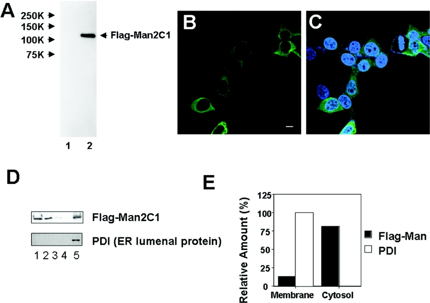
Figure 1. Expression of human mannosidase in mammalian cells.
(A) Western blotting analysis of the cytosolic fraction. Lane 1, HEK-293 cells with mock plasmid; lane 2, HEK-293 cells with FLAG–Man2C1 expression plasmid. Stained with anti-FLAG antibody. (B and C) FLAG–Man2C1 was immunostained (green) with an anti-FLAG antibody. In (C), nuclei were stained with DAPI (blue). Bar=10 μm. (D) Subcellular fractionation of FLAG–Man2C1. Lane 1, cytosolic fraction; lanes 2–4, first, second and third wash of the membrane fraction; lane 5, membrane fraction. PDI is a marker for ER luminal protein. (E) Distribution of FLAG–Man2C1. The results were obtained by quantitation of results in (D).

